 English
 한국어
한국어 français
 Deutsch
 Español
 русский
 português
 العربية
 ไทย
ไทย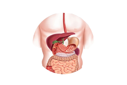
Address
1st & 2nd Floor, 10 Building, 18 Huashan Rd., Changzhou, Jiangsu province, China

ERCP, or endoscopic retrograde cholangiopancreatography, is a non-invasive or minimally invasive diagnostic and therapeutic method for hepatobiliary pancreatic system diseases that has been around for 50 years. In 1968, researchers at the University of Washington performed the first cannulation of the duodenal papilla using a side-viewing fiberoptic duodenoscope. The scope had an angle of 90° between the objective lens and the eyepiece, which was ideal for observing the papilla on the wall of the duodenum and for cannulation under direct view. They assembled a set of instruments for endoscopic insertion into the bile duct and pancreatic duct and placed a catheter on the Eder-type fiberoptic duodenoscope. The catheter was placed through a balloon dilatation of the papilla of Vater under direct view, and the clinical application of this technique was reported for the first time, but the success rate of cannulation was only 25% at that time. In 1970, Japanese scholars further studied and improved the technique, and reported 60 successful medical ERCP procedures, which gradually became widely used in clinical practice around the world and became an important diagnostic technique for biliary and pancreatic diseases. It was these pioneers' continuous innovation and exploration that laid a solid foundation for ERCP.
The early medical ERCP technique was used only as an auxiliary diagnostic technique. Operators injected contrast agents to understand the disease conditions inside the biliary and pancreatic ducts, and because CT technology was still immature at that time, medical ERCP had a high clinical value in diagnosis. Subsequently, clinical doctors began to attempt therapeutic operations using ERCP. In 1970, Professor Soehendra of Germany designed the first plastic biliary stent. Endoscopic retrograde biliary drainage (ERBD) was also reported for the first time for the treatment of obstructive biliary diseases. Subsequently, endoscopic sphincterotomy (EST) was developed, which solved the problem of the barrier posed by the natural barrier of the Oddi sphincter to the entry and exit of various operative instruments. ERCP gradually became a first-line treatment for common bile duct stones.
In 1975, Japanese scholars first attempted nasobiliary drainage through a duodenoscope. In 1977, Web and Classen conducted a large-sample clinical study of endoscopic nasobiliary drainage (ENBD), confirming its effectiveness in the treatment of acute suppurative cholangitis. In 1983, Dr. Seigle used a plastic stent to treat obstructive pancreatic diseases for the first time. In 1985, Carrasco first attempted to place a metal stent used for blood vessels in the bile duct, opening up the application of expandable metal stents in the treatment of biliary strictures. It was then gradually widely used for the treatment of malignant biliary obstruction worldwide. Subsequently, expandable metal stents used to treat pancreatic duct obstruction caused by pancreatic head cancer received increasingly widespread clinical application.
With the continuous development of endoscopic equipment, various operations derived from traditional medical ERCP have become more and more diverse. In 1971, endoscopic pancreatic juice collection and molecular biology examination techniques emerged, greatly improving the accuracy of differential diagnosis of pancreatic diseases. The emergence of biliary and pancreatic duct brushing in 1975 further improved the diagnostic sensitivity of biliary and pancreatic diseases.
In the 1980s and 1990s, endoscopic papillary balloon dilation (EPBD), peroral cholangioscopy, peroral pancreatoscopy, endoscopic retrograde cholecystocholangiography and stent placement, and other techniques emerged. In recent years, with the development of intraductal ultrasound technology, intraductal ultrasonography of the pancreatobiliary duct has been performed, which overcomes the defect of medical ERCP being able to observe only the shape of the duct and unable to observe intraluminal or intramural lesions.
The submucosal endoscope visualization system, which was introduced in 2007, can enter the bile and pancreatic duct cavities, observe the surface structure of the stenotic section, and perform biopsies under direct view, greatly improving the positive rate of biopsies and allowing accurate diagnoses of rare biliary and pancreatic duct lesions. Through continuous recognition, understanding, and exploration of diseases, as well as continuous research and innovation of endoscopic diagnostic and therapeutic techniques, new examination and treatment methods have continued to emerge, and the new generation of medical ERCP diagnostic and therapeutic technologies such as confocal laser endomicroscopy, photodynamic therapy, and endoscopic radiofrequency ablation have gradually been applied in clinical practice. Looking ahead, ERCP will continue to play a pivotal role in the diagnosis and treatment of hepatobiliary and pancreatic diseases.